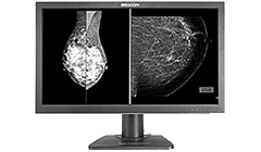
 Diagnostic Display



Why Mini LED Is Unsuited for Diagnostic Radiology Monitors
 2026-10-23
In recent years, Mini LED backlight technology has gained widespread adoption in consumer electronics and certain medical fields—such as surgery and endoscopy—owing to its high brightness, superior contrast, and precise backlight control. These technological advantages effectively meet demands for high brightness and dynamic image viewing.
However, diagnostic radiology imaging has distinct requirements that set it apart from standard visual displays. When physicians read medical images such as CT scans, MRIs, digital radiography (DR), and mammograms, they are not primarily looking for brighter visuals or deeper blacks. Instead, they require subtle grayscale differences to be presented accurately, stably, and consistently to precisely identify pathological features.
Therefore, for Mini LED technology solutions that utilize dynamic local dimming, it is crucial to evaluate how well they align with the rigorous demands of diagnostic radiology. The advantages of high brightness and high contrast do not automatically translate into superior diagnostic display performance. This discrepancy stems from a structural misalignment between the imaging mechanism of Mini LED and the clinical requirements of diagnostic radiology.
Starting from display technology principles and combining the DICOM standard with clinical reading scenarios, this article examines the technical root causes of this mismatch.
Part 1: Technical Characteristics of Mini LE

The core principle of Mini LED is replacing the single backlight module of traditional LCDs with hundreds to thousands of independently controlled LED zones. This enables individual or grouped control of backlight brightness across different areas of the screen. Through local dimming algorithms, when high contrast areas appear on the screen, the system increases the backlight brightness in bright regions while decreasing it in dark regions, thereby enhancing dynamic contrast and dark-field performance. This technology is particularly well-suited for high visual impact scenarios such as HDR video, movies, and gaming.
However, Mini LED increases the granularity of backlight control; it does not mean that the LCD pixels themselves possess independent light-emitting capabilities. Backlight zones and liquid crystal pixels do not maintain a one-to-one correspondence. When a single backlight zone contains both bright and dark pixels simultaneously, the system must still compromise between the two. This limitation forms a key technical challenge that must be thoroughly evaluated when local dimming technology is applied to high-precision medical imaging displays.
Part 2: Core Requirements of Diagnostic Radiology Displays: Accurate Reproduction
Diagnostic radiology monitors are primarily used for the clinical interpretation of medical images. For the observation of micro-lesions, low-contrast tissue, subtle density variations, and lesion margins, a display must accurately reproduce image information to avoid introducing artificial brightness shifts or spatial interference. Its technical index system and evaluation standards differ fundamentally from consumer-grade displays, as reflected in four key dimensions:
1. Grayscale Accuracy: Compliance with DICOM GSDF Display Requirements

Medical images typically contain rich grayscale information. Taking CT scans as an example, physicians identify lesions based on density differences between tissues; in chest X-rays and mammography, minute grayscale changes can also impact lesion identification and morphological interpretation.
DICOM PS3.14 defines the Grayscale Standard Display Function (GSDF) to standardize the grayscale-to-luminance response of display systems. This ensures that brightness variations across different grayscale levels align closely with human visual perception characteristics, serving as one of the key technical foundations for medical image display consistency.
The risk associated with Mini LED displays utilizing dynamic local dimming is that backlight luminance dynamically interferes based on image content and local brightness distribution. If the same grayscale value corresponds to different actual luminance levels under varying image conditions, it can compromise grayscale consistency. When algorithms judge that a specific region "should be brighter" or "should be darker" based on content, the actual luminance mapping deviates from the DICOM standard. For diagnostic scenarios requiring precise density evaluation, this uncertainty is unacceptable.
2. The Halo Effect: Impeding the Observation of Micro-Structures and Low-Contrast Lesions

Mini LED local dimming presents a physical-level contradiction: backlights can be controlled by zones, but liquid crystal pixels cannot. Because a single backlight zone typically covers multiple LCD pixels, when high-brightness structures and dark backgrounds fall into the same or adjacent zones, the backlight cannot independently adjust to the exact luminance needs of every pixel. Combined with optical diffusion and other factors, brightness spreading or edge halos may occur around high-brightness objects.
In radiological imaging, physicians frequently need to observe small, low-contrast structures, such as:
- Chest CT / X-rays: Subtle differences between ground-glass nodules and surrounding lung tissue, as well as fine grayscale changes across lung fields, the mediastinum, and other structures.
- Mammography: The morphology and distribution of microcalcifications and their relationship with surrounding tissue.
If local dimming alters the brightness relationships near a lesion, it can increase the difficulty of detail observation. Consequently, halos and their impact on low-contrast details can directly obscure or blur the boundaries of micro-lesions, potentially leading to interpretation issues in early-stage lesion morphology.
3. The Contradiction Between Static Reading and Dynamic Dimming

Diagnostic radiology reading relies primarily on static images. Physicians repeatedly observe, compare, and measure a single image, with viewing times for one image lasting anywhere from tens of seconds to several minutes. Mini LED local dimming algorithms are optimized for dynamic content and can introduce subtle brightness fluctuations in static images. Although minor, these fluctuations can cause visual fatigue and distraction during prolonged reading sessions.
More importantly, diagnostic radiology demands far higher full-screen luminance uniformity than peak brightness. The core metric for a diagnostic monitor is whether "the brightness in every single corner of the screen is consistent and stable," rather than "how high the maximum nit output can be." Mini LED's dense backlight zones actually increase the complexity of uniformity control.
4. Conflict Between Calibration Determinism and Algorithmic Indeterminacy

Diagnostic radiology monitors require regular DICOM GSDF calibration to ensure the determinism and traceability of grayscale output. Traditional LCDs combined with high-precision backlight control provide deterministic, predictable, and calibratable luminance output. Mini LED's local dimming algorithms introduce a dynamic variable: the same grayscale value may correspond to different actual luminance outputs depending on the image content. This poses a fundamental challenge to calibration, as the calibration target is no longer just the display hardware itself, but a dimming algorithm that constantly shifts with the image content.
Conclusion: Mini LED Is Unsuited for Diagnostic Radiology Scenarios
For diagnostic radiology applications, a brighter image or deeper black levels do not equate to more accurate lesion visualization. Medical imaging display requires DICOM GSDF calibration, grayscale linearity, full-screen uniformity, and long-term stability—prioritizing the precise and stable presentation of every subtle grayscale difference in medical images over visual impact.
Traditional LCDs paired with stable backlight control and comprehensive calibration systems remain a mature, reliable, and preferred technology pathway.
Currently, Mini LED technology in the medical field is primarily deployed in surgical display scenarios. For diagnostic radiology, its clinical applicability requires careful evaluation. Technology selection in the medical display industry must respect regulatory requirements, ensuring accuracy, stability, and consistency to prevent the misdiagnosis or missed diagnosis of early-stage lesions.
 Back to list
News Recommendation